The International Elbow Working Group's four-grade classification system forms the foundation of elbow dysplasia screening worldwide. Established in 1990 and refined through subsequent revisions, this protocol aims to standardize assessment across international borders, enabling meaningful comparison of screening data and facilitating informed breeding decisions. Yet the apparent simplicity of a 0-3 scale masks considerable complexity in interpretation, and understanding what each grade represents pathologically is essential for both veterinary professionals and breeders evaluating certification results.
Historical Development of the IEWG System
The International Elbow Working Group formed in 1989 when veterinary radiologists from 14 countries recognized that elbow dysplasia screening lacked the standardization achieved for hip dysplasia. Each nation employed different evaluation criteria, making cross-border comparison of breeding stock impossible and impeding epidemiological research.
The founding IEWG meeting in Stockholm established core principles: a common terminology for the three ED components (FCP, OCD, UAP), minimum radiographic requirements, age standards for official certification, and a grading scale reflecting disease severity. The initial 1990 guidelines underwent significant revision in 2001, adding clarification for borderline cases and introducing the concept of elbow incongruity assessment.
IEWG Member Organizations
The IEWG includes representatives from over 30 national kennel clubs and veterinary organizations. Member bodies agree to apply IEWG principles, though implementation varies. The FCI (Federation Cynologique Internationale) adopted IEWG protocols for member countries, while the OFA developed parallel standards for North America that align closely but not identically with IEWG guidelines.
The Four-Grade System Explained
IEWG grading assesses both primary lesions (FCP, OCD, UAP) and secondary osteoarthritis, assigning the higher of the two findings as the final grade. This dual assessment recognizes that some dogs develop significant secondary osteoarthritis from subtle primary lesions, while others show obvious primary lesions with minimal secondary changes.
Grade 0
Normal - No evidence of ED
Grade 1
Mild ED - Minimal changes
Grade 2
Moderate ED - Obvious changes
Grade 3
Severe ED - Advanced disease
Grade 0: Normal Elbows
A Grade 0 classification indicates that no radiographic evidence of elbow dysplasia or secondary osteoarthritis is present. The joint surfaces appear smooth and congruent, with no osteophyte formation, sclerosis, or visible primary lesions.
Radiographic Criteria for Grade 0
- Anconeal process fully fused with smooth margins
- Medial coronoid process of normal shape without blunting or fragmentation
- Humeral condyle with smooth articular surface, no defects
- No osteophytes on anconeal process, radial head, or epicondyles
- No subchondral sclerosis of the trochlear notch
- Joint space uniform, no evidence of incongruity
Grade 0 Is Not Genetic Clearance
A Grade 0 phenotype does not guarantee absence of ED-predisposing genes. Dogs with Grade 0 elbows can and do produce affected offspring, particularly when mated to partners with unfavorable pedigrees. Breeding decisions should consider family history, not just individual screening results. Among Grade 0 x Grade 0 matings, offspring ED rates range from 5-15% depending on extended family history.
Grade 1: Mild Elbow Dysplasia
Grade 1 represents minimal radiographic changes that may be difficult to distinguish from normal variation. This borderline category generates the most inter-observer disagreement, with studies showing only 72% agreement between certified evaluators on Grade 0 versus Grade 1 classification.
Radiographic Criteria for Grade 1
- Osteophytes less than 2mm on the anconeal process
- Mild osteophytes on the dorsal aspect of the radial head
- Slight sclerosis of the trochlear notch base
- Subtle irregularity of the medial coronoid apex
- Very mild joint incongruity (subjective assessment)
- No visible primary lesion (FCP, OCD, UAP fragment)
The Grade 1 Dilemma
Grade 1 presents the most challenging classification because it encompasses dogs with genuine early-stage ED and dogs with borderline-normal anatomy misinterpreted due to positioning or image quality. Many breed clubs prohibit Grade 1 breeding entirely, while others permit breeding only to Grade 0 partners. The appropriate response depends on breed baseline prevalence and the individual dog's pedigree analysis.
Clinical Significance of Grade 1
Dogs with Grade 1 ED are typically clinically sound at the time of screening. Long-term studies suggest that Grade 1 dogs have elevated risk of developing symptomatic osteoarthritis by middle age compared to Grade 0 dogs, though many remain comfortable throughout life with appropriate management. Heritability of Grade 1 is lower than Grade 2-3, suggesting greater environmental contribution to these minimal changes.
Grade 2: Moderate Elbow Dysplasia
Grade 2 indicates definite elbow dysplasia with obvious radiographic changes. Dogs in this category have unequivocal pathology that affects both their clinical prognosis and breeding suitability.

Radiographic Criteria for Grade 2
- Osteophytes 2-5mm on anconeal process or radial head
- Obvious sclerosis of the trochlear notch
- Blunting or remodeling of the medial coronoid process
- Visible but undisplaced UAP or OCD lesion
- Moderate incongruity visible on standard views
- Evidence of cartilage erosion or subchondral bone exposure
Clinical and Breeding Implications
Grade 2 dogs often show subtle lameness or stiffness, particularly after exercise or upon rising from rest. Progressive osteoarthritis is expected, and most Grade 2 dogs will require ongoing management including weight control, activity modification, and possibly pharmaceutical support.
From a breeding perspective, Grade 2 represents a breeding exclusion across virtually all responsible breeding programs. The heritability of Grade 2 ED is substantial (0.35-0.45), and breeding affected dogs perpetuates the condition regardless of partner quality.
Grade 3: Severe Elbow Dysplasia
Grade 3 indicates advanced elbow dysplasia with marked radiographic changes and typically significant clinical impact. Dogs with Grade 3 ED have substantial pathology requiring active management.
Radiographic Criteria for Grade 3
- Osteophytes greater than 5mm
- Displaced or fragmented UAP clearly visible
- Large OCD lesion with flap detachment
- Advanced secondary osteoarthritis
- Joint deformity or severe incongruity
- Eburnation (bone-on-bone contact changes)
Clinical Presentation
Most Grade 3 dogs demonstrate obvious lameness, reduced range of motion, and joint effusion on examination. The condition significantly impacts quality of life, and surgical intervention is typically indicated. Even with treatment, lifelong osteoarthritis management is necessary.
Absolute Breeding Exclusion
No justification exists for breeding Grade 3 dogs under any circumstances. These dogs carry significant genetic load for ED, and breeding them contributes to population-level disease perpetuation. Breed clubs universally prohibit Grade 3 breeding, and ethical breeders should refuse stud services or breeding involving Grade 3 individuals regardless of other qualities.
Grading the Components: FCP, OCD, UAP
When specific primary lesions are identified, IEWG protocols assign grades based on lesion characteristics:
| Lesion | Grade 2 Criteria | Grade 3 Criteria |
|---|---|---|
| FCP | Coronoid blunting, sclerosis, suspected fragment | Clearly displaced fragment, severe sclerosis |
| OCD | Visible defect on humeral condyle | Large defect with detached flap or joint mice |
| UAP | Visible lucent line, process not displaced | Displaced or rotated unfused anconeal |
Primary lesions automatically result in Grade 2 or 3 classification regardless of secondary OA severity
Incongruity Assessment
The 2001 IEWG revision added formal recognition of elbow incongruity, though its inclusion in grading remains inconsistent across member organizations. Incongruity refers to radio-ulnar step defects that alter joint contact mechanics, potentially contributing to all three classical ED lesions.
Short Radius Incongruity
The radial head sits below the level of the ulnar articular surface. This shifts weight-bearing posteriorly, increasing anconeal loading. Associated with UAP development.
Short Ulna Incongruity
The ulnar articular surface sits below the radius. The medial coronoid bears excessive load. Strongly associated with FCP development.
Current IEWG guidelines suggest noting incongruity presence but do not specify separate grading criteria. Some national schemes assign Grade 1 for mild incongruity without other changes, while others only grade incongruity when accompanied by osteophyte formation.
Inter-Observer Variability
Studies consistently demonstrate significant disagreement between certified evaluators, particularly at the Grade 0/1 boundary. Lang et al. (2007) found 72% agreement for this distinction among IEWG-certified screeners. Agreement improves for higher grades, reaching 90%+ for Grade 2/3 differentiation.
| Classification Decision | Observer Agreement | Main Sources of Disagreement |
|---|---|---|
| Grade 0 vs Grade 1 | 70-75% | Subtle osteophytes, sclerosis interpretation |
| Grade 1 vs Grade 2 | 80-85% | Osteophyte size measurement, coronoid assessment |
| Grade 2 vs Grade 3 | 90-95% | Minor disagreements on lesion displacement |
Implications for Breeders
This variability has practical implications for breeding decisions. A dog graded borderline Grade 1 by one evaluator might receive Grade 0 from another. When results seem inconsistent with clinical examination or family history, consider requesting re-evaluation by a different certified screener before making irreversible breeding decisions.
Reporting Final Grades
IEWG protocol assigns the final grade as the worse of the two elbows. A dog with Grade 0 left elbow and Grade 1 right elbow receives an overall Grade 1 classification. This approach ensures that breeding decisions account for any disease present.
The BVA/KC scheme notably diverges from this approach, reporting individual elbow scores (e.g., 0:1) along with a total. This provides more information for pedigree analysis, as a 0:0 parent is likely lower genetic risk than a 0:2 parent, even though both would receive Grade 2 under IEWG reporting.
Example IEWG Certificate Statement
Elbow Grade: 1 (ED 1) Right elbow: Grade 0 | Left elbow: Grade 1 | Final classification based on worse elbow
Minimum Standards for Valid Evaluation
IEWG guidelines specify technical requirements for certification-quality radiographs:
- Minimum age: 12 months (some registries require 24 months for official certification)
- Required views: Mediolateral flexed and craniocaudal (extended ML recommended)
- Positioning: Minimal rotation; epicondyles should superimpose on ML view
- Image quality: Adequate exposure allowing bone detail assessment without overexposure
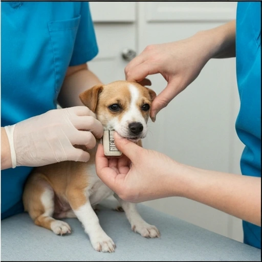
- Identification: Permanent ID (microchip or tattoo) linked to radiograph
Related Database Resources
- Comparing Grading Systems - IEWG vs OFA vs BVA/KC protocols
- Radiographic Diagnosis - Imaging techniques for accurate grading
- Breeding Decisions - Translating grades into breeding recommendations
- Certification Requirements - What breeders need for registration
Conclusion
The IEWG grading system provides a standardized framework for elbow dysplasia assessment, enabling international comparison and informing breeding decisions. Understanding what each grade represents pathologically, recognizing the inherent limitations of inter-observer variability, and integrating individual results with pedigree analysis allows breeders to make informed decisions that contribute to population-level improvement in elbow health.